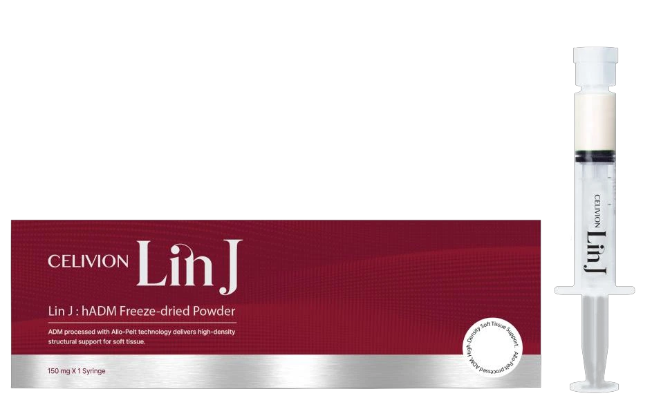
What is Celivion Lin J?
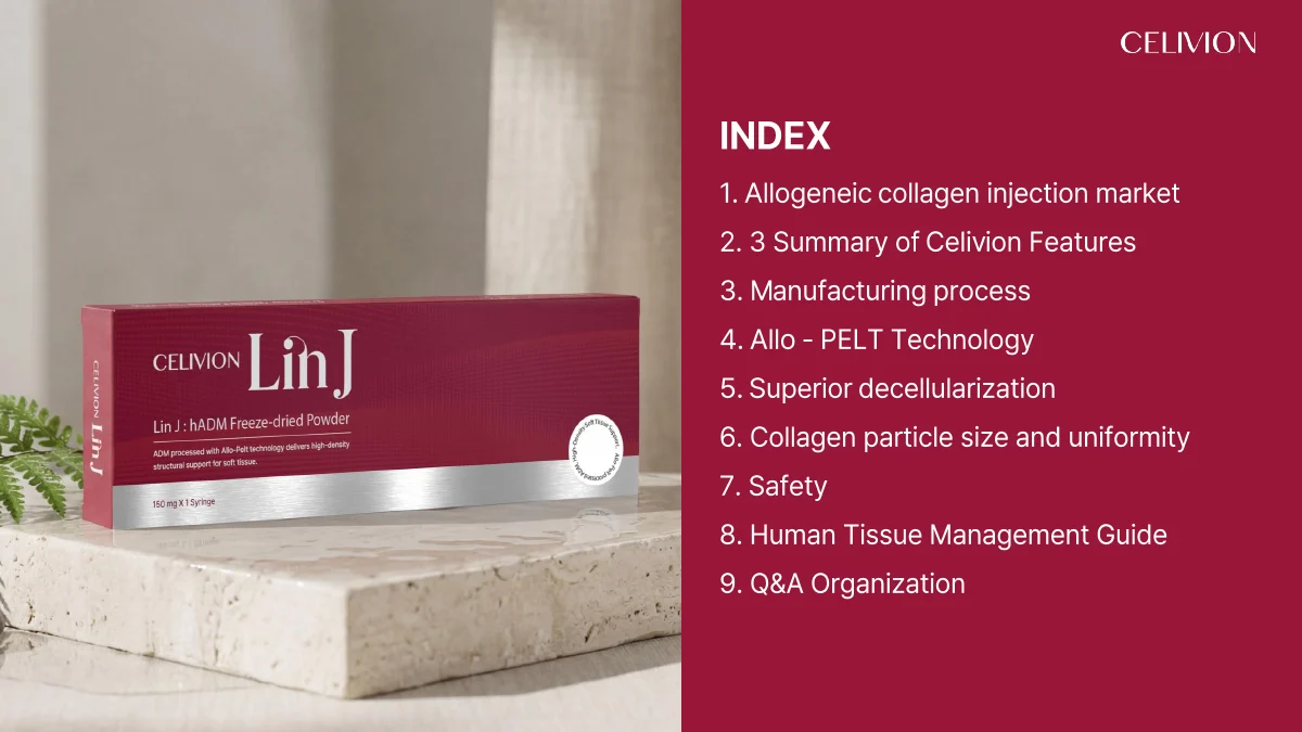
A product based on human acellular dermal matrix (hADM), processed into fine particles. The technical dossier describes its material structure, processing and laboratory results.
Technical library / Source edition 042326L
Explore the structure.
Read the evidence.
Explore Celivion Lin J hADM, Allo-PELT processing, ECM composition, particle size, DNA and laboratory results, with source pages and technical questions.
Values reported in this technical dossier · pp. 14, 18, 25
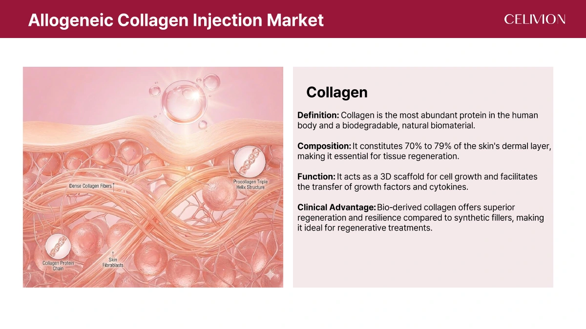
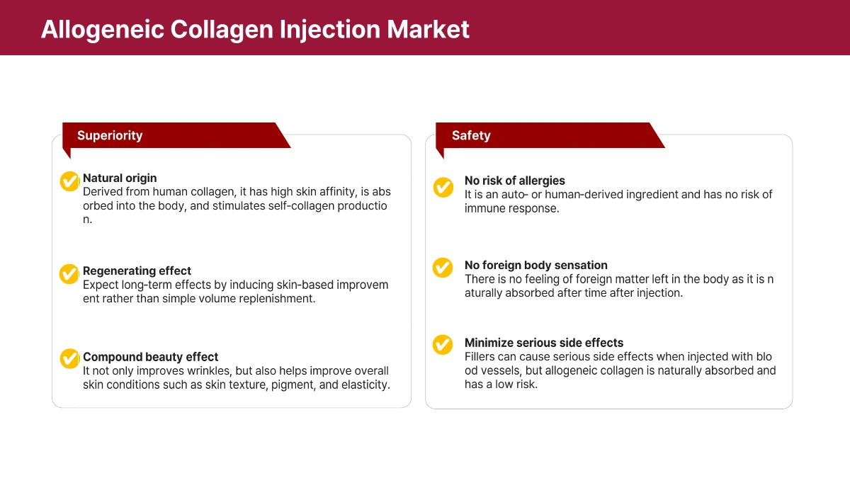
Celivion Lin J is based on human acellular dermal matrix (hADM). Processing removes cellular components while retaining extracellular matrix (ECM), including collagen. hADM is a tissue matrix; it is not a living-cell product.
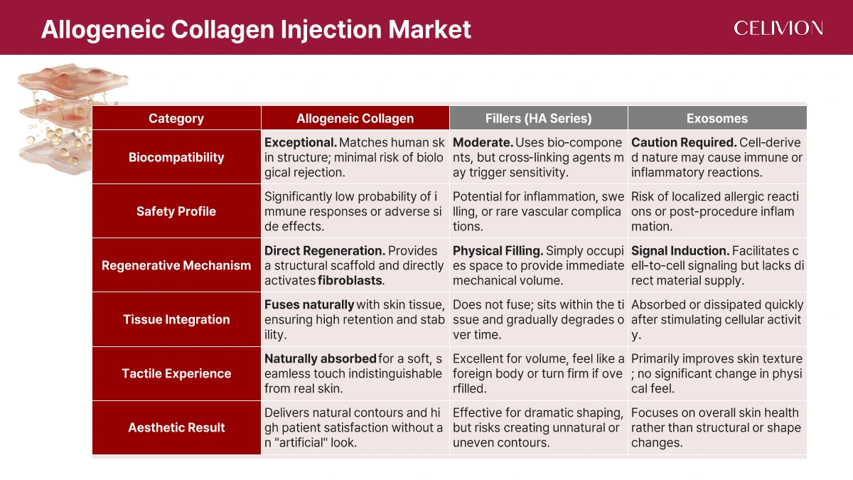
The dossier distinguishes three material approaches: an allogeneic collagen/ECM scaffold, hyaluronic acid (HA) filling, and exosome-associated signalling. These describe different material concepts, not a head-to-head Celivion Lin J clinical trial.
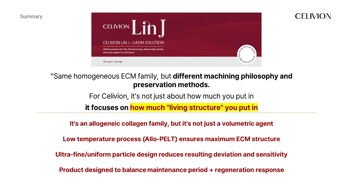
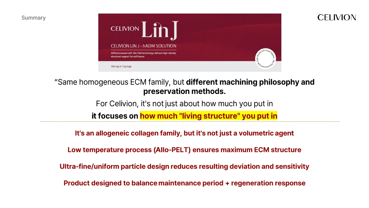
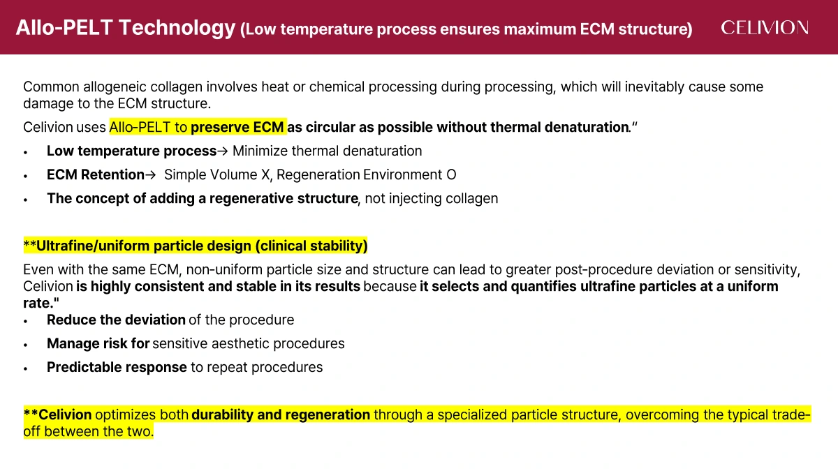
Celivion Lin J’s three design priorities are low-temperature ECM preservation, fine and uniform particle processing, and the relationship between material persistence and tissue response.
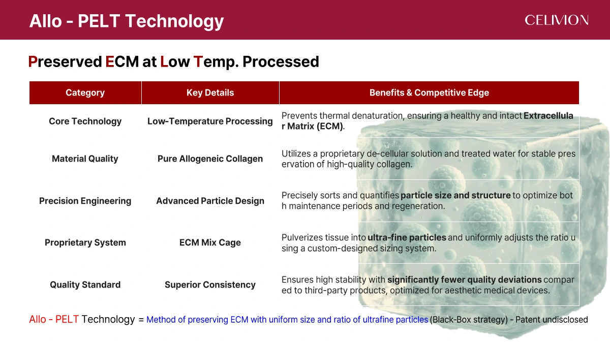
Allo-PELT stands for Preserved ECM at Low Temperature Processing. The source describes a staged low-temperature circulation process, decellularization solution and treated water, followed by particle sizing and ratio control using an “ECM Mix Cage” system.
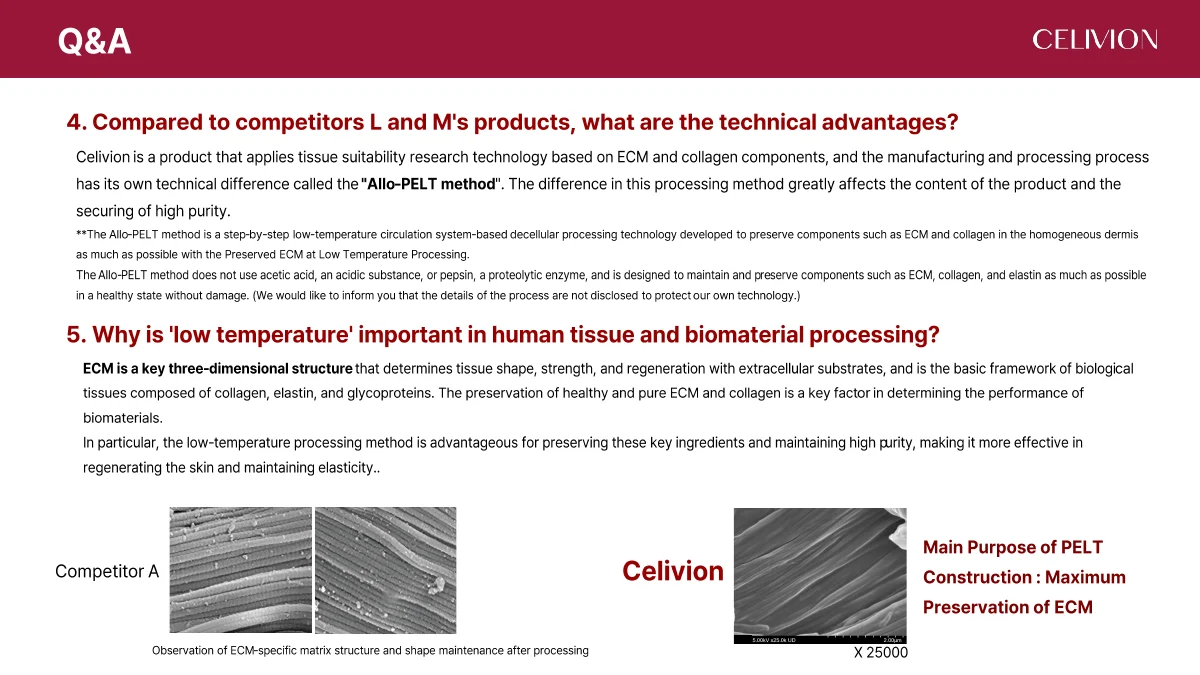
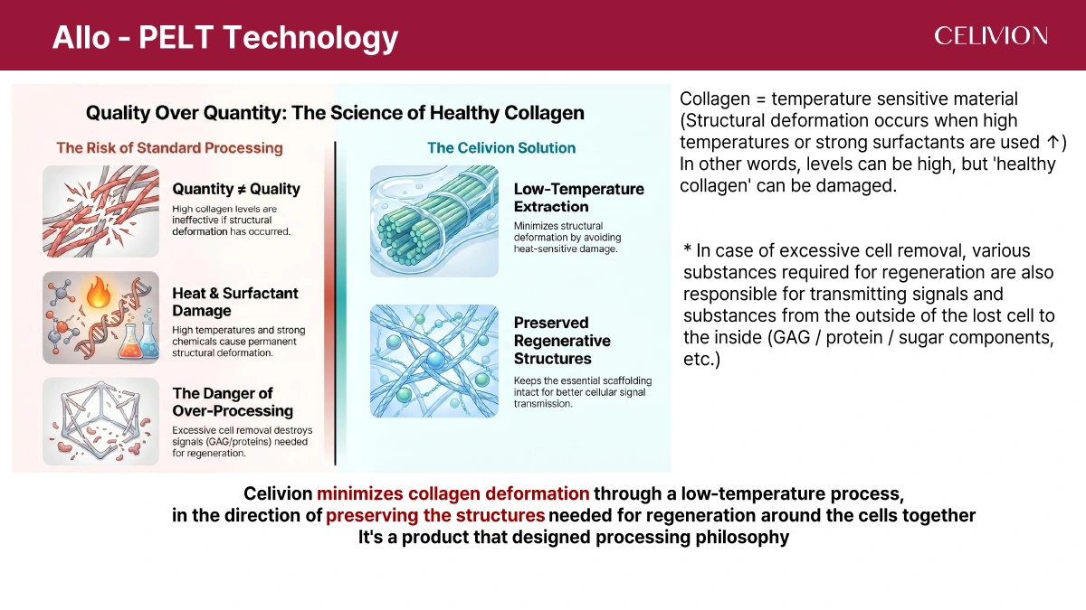
The Celivion dossier states that this process does not use acetic acid or pepsin. Its design objective is to retain collagen, elastin and other ECM components while limiting thermal deformation. Detailed proprietary process parameters are not disclosed.
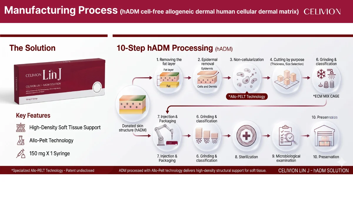
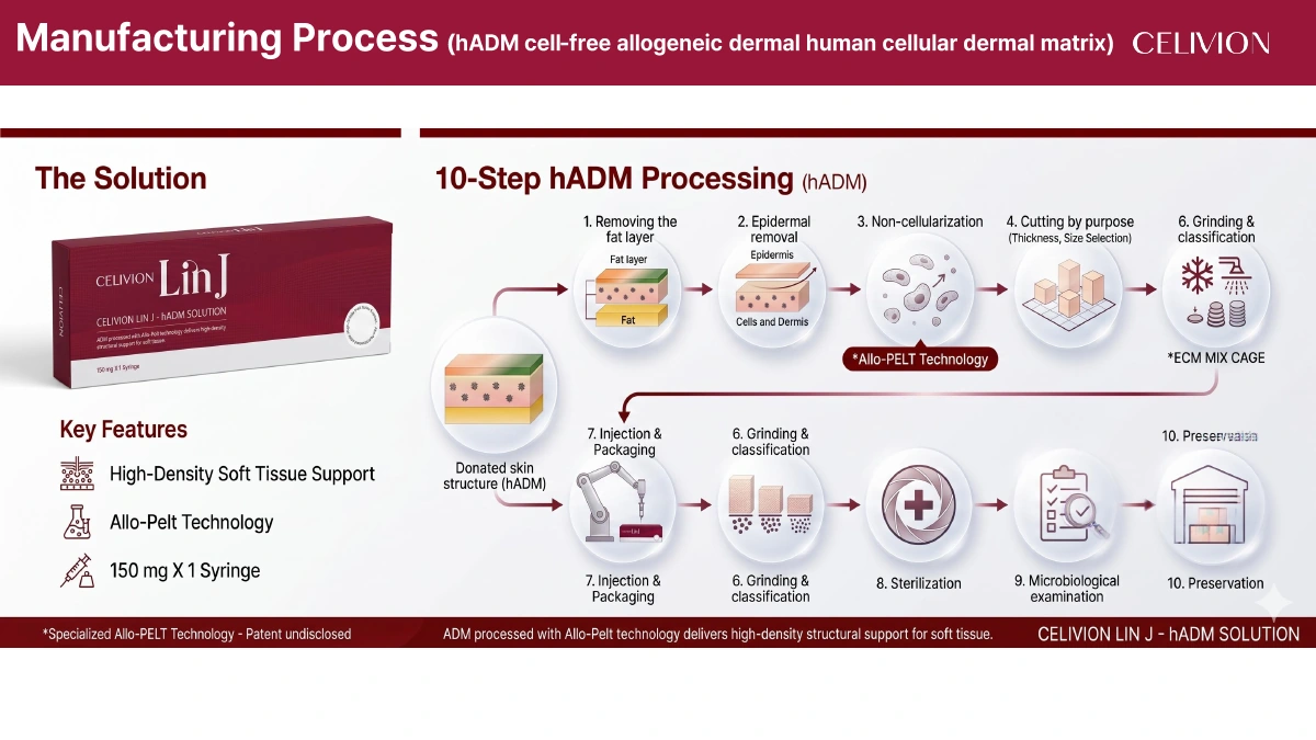
Source page 8 presents the manufacturing flow. Pages 9 and 34 explain the processing approach; the source flow diagram remains available below.
The manufacturing diagram names fat-layer removal, epidermal removal, decellularization, purpose-specific cutting, grinding and classification, filling and packaging, sterilization, microbiological examination and preservation. The labels follow the source; they are a manufacturing overview, not an operating protocol.
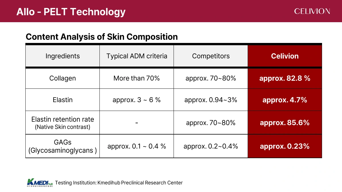
The Celivion source reports the following composition analysis from Kmedihub Preclinical Research Center. Values are reproduced as given, including approximate values and the unnamed comparator column. These are source-reported material measurements, not clinical outcome percentages.
| Component | Typical ADM criteria | Comparators | Celivion |
|---|---|---|---|
| Collagen | >70% | ≈70–80% | ≈82.8% |
| Elastin | ≈3–6% | ≈0.94–3% | ≈4.7% |
| Elastin retention vs. native skin | — | ≈70–80% | ≈85.6% |
| GAGs | ≈0.1–0.4% | ≈0.2–0.4% | ≈0.23% |
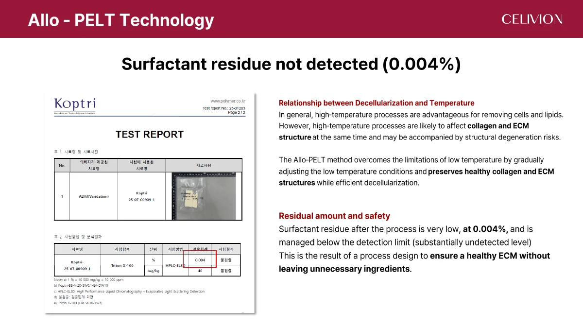
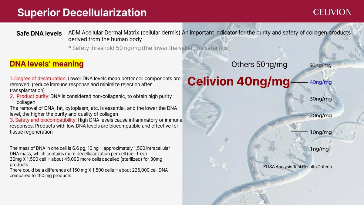
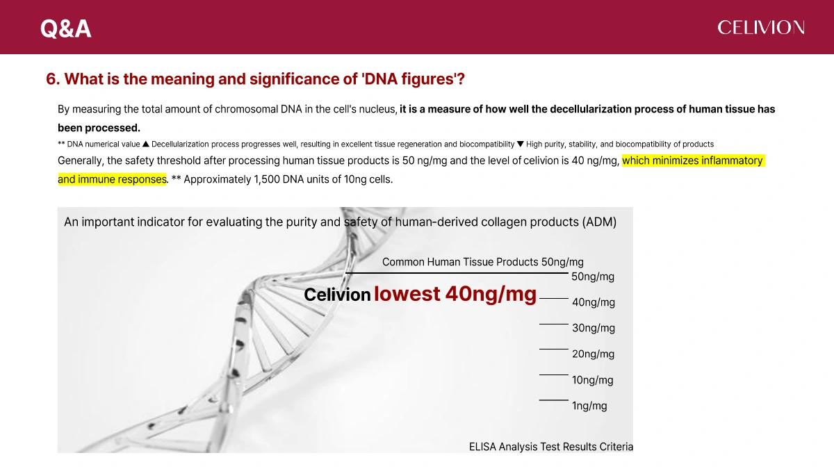
The dossier reports residual DNA of 40 ng/mg for Celivion, alongside a 50 ng/mg reference threshold. Residual DNA is one indicator of cellular-material removal and does not by itself establish overall product safety.
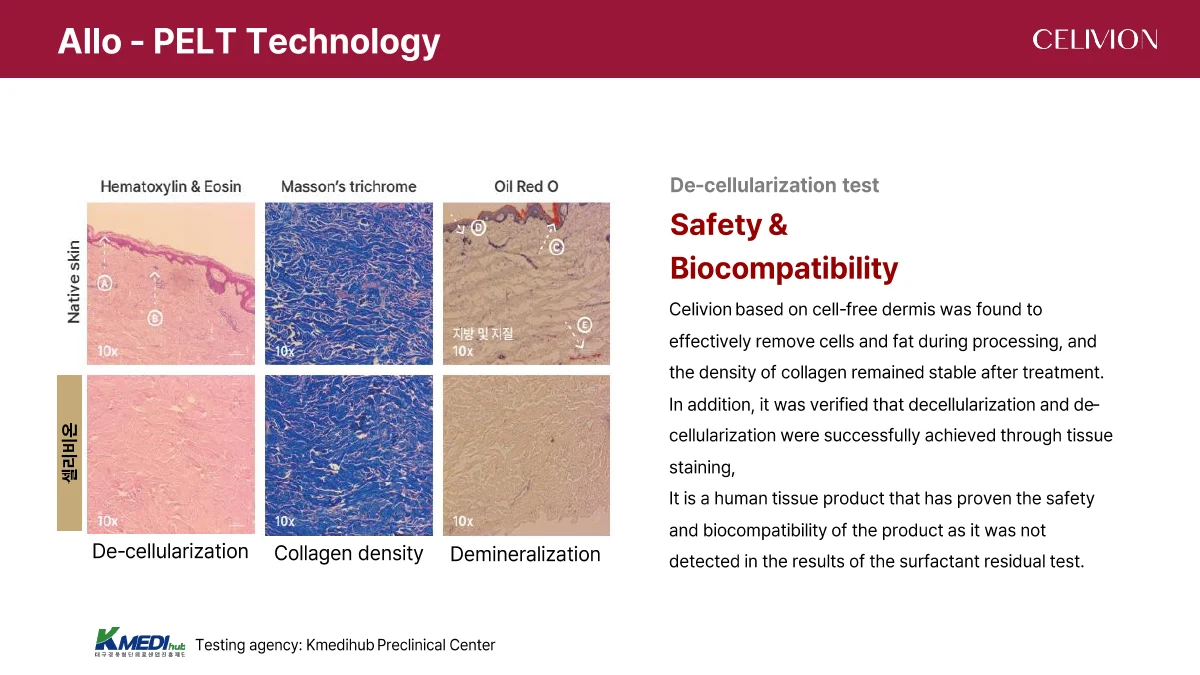
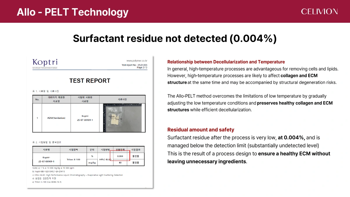
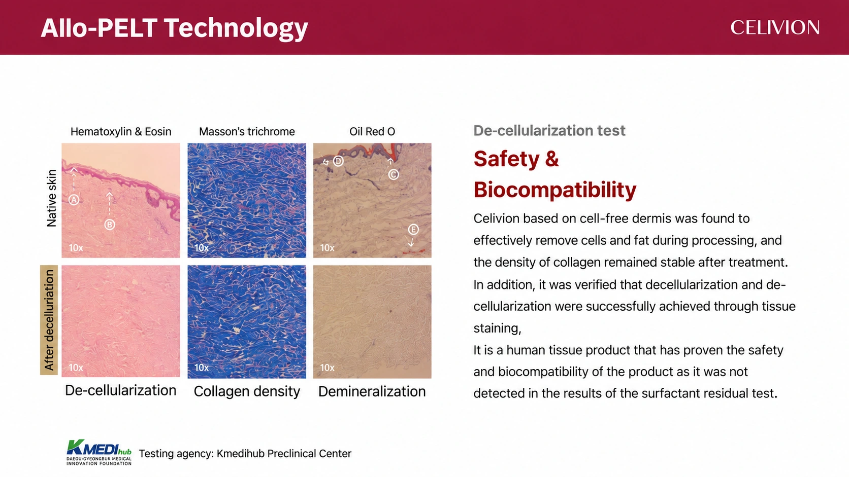
Tissue staining is used to examine cellular removal, collagen density and lipid removal. Source page 12 reports 0.004% surfactant residue while also using “not detected” wording. This page preserves the numerical result without treating it as zero; the detection limit and complete test report are needed to interpret that wording.
Testing institution named for the histology and residual analysis: Kmedihub Preclinical Center.
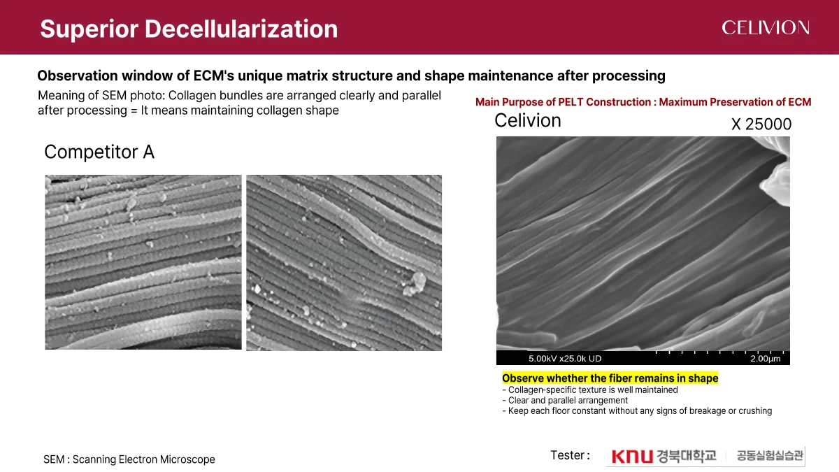
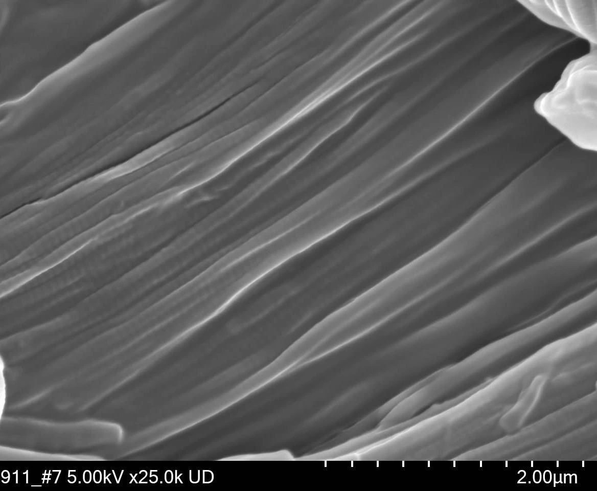
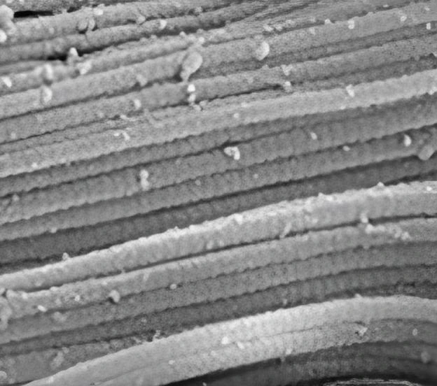
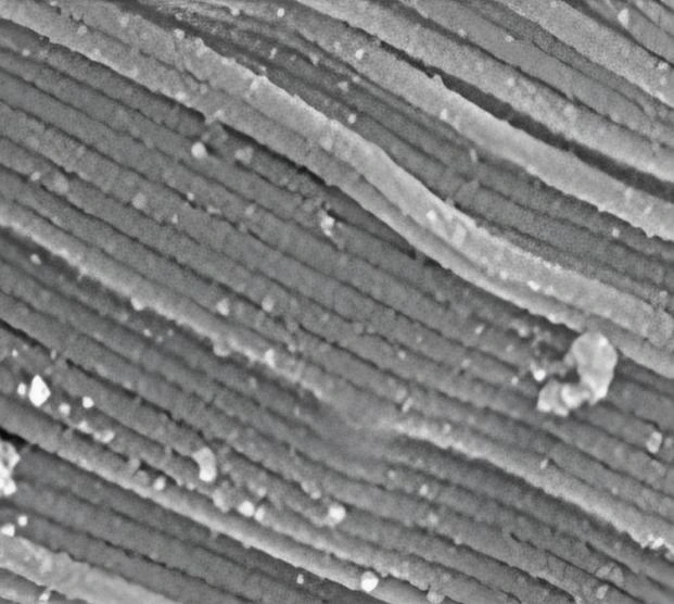
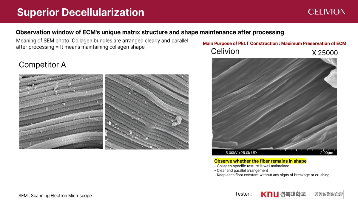
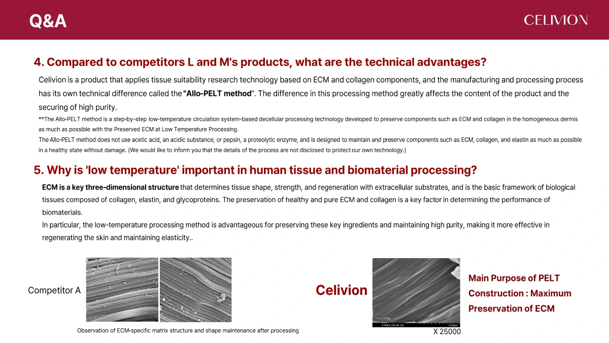
Scanning electron microscopy (SEM) examines collagen fibre shape and arrangement after processing. The source shows Celivion and comparator A, with a ×25,000 label on the Celivion image. These are morphological observations.
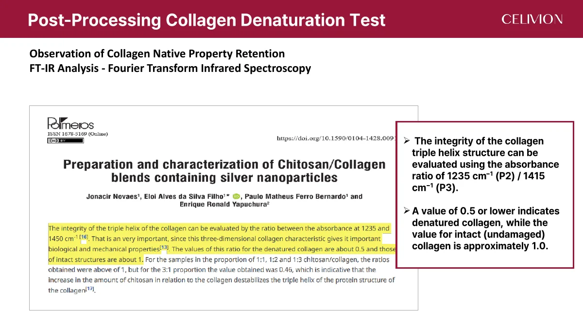
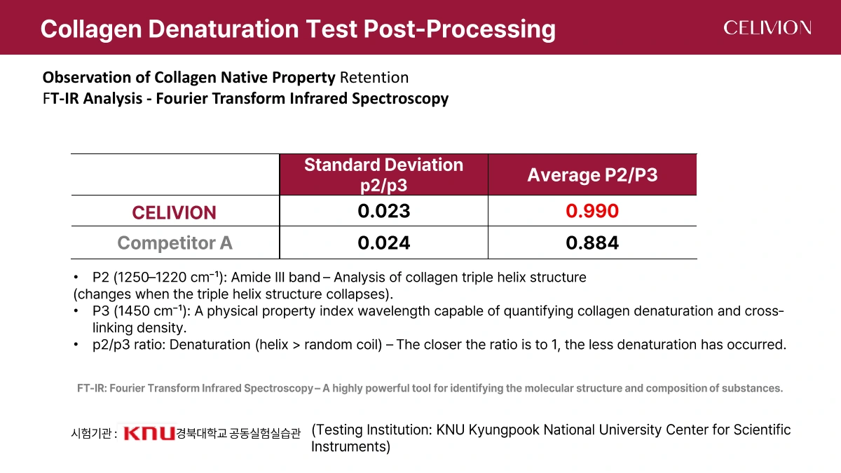
FT-IR examines molecular structure through infrared absorption. The source reports a mean P2/P3 ratio of 0.990 for Celivion and 0.884 for comparator A, with standard deviations of 0.023 and 0.024. Testing institution: Kyungpook National University Center for Scientific Instruments.
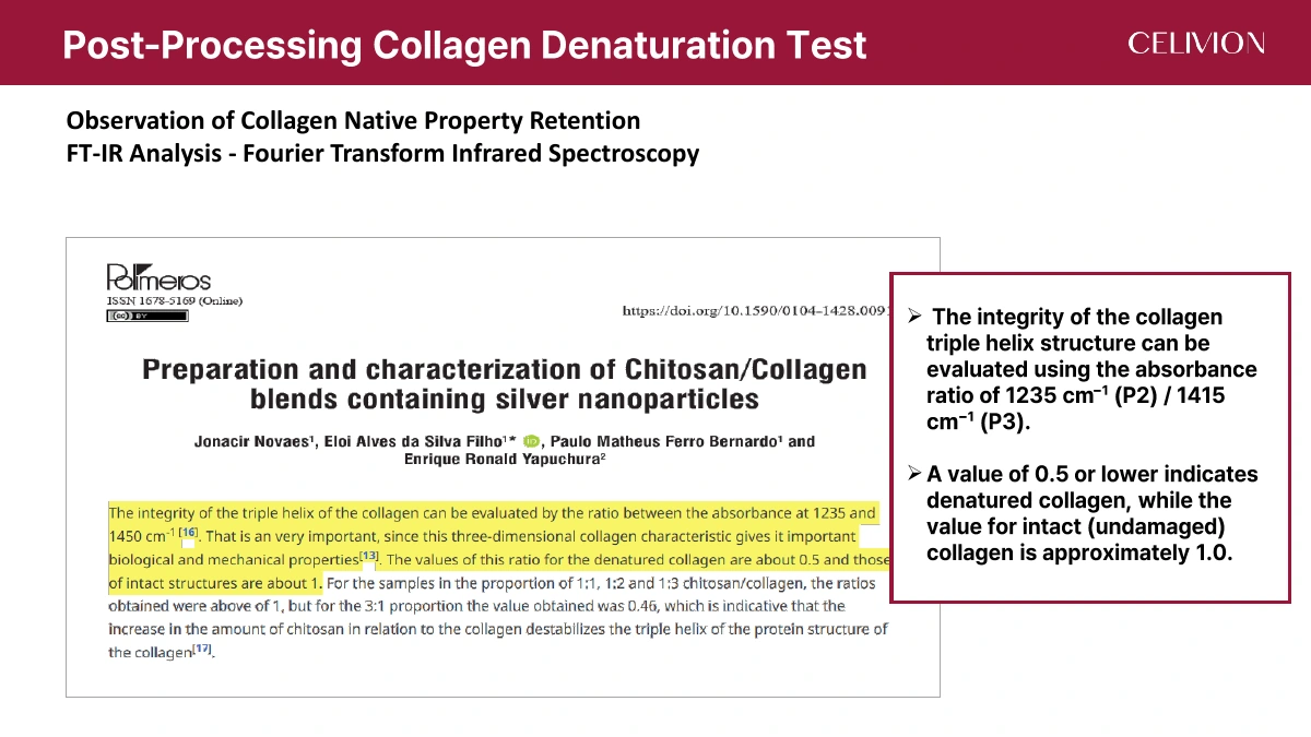
Page 16 labels P2 at 1250–1220 cm⁻¹ and P3 at 1450 cm⁻¹. Page 17 cites another analysis using 1235/1415 cm⁻¹, with approximately 1.0 for intact collagen and ≤0.5 for denatured collagen. The two wavelength descriptions are retained separately.
| Material | P2/P3 mean | Standard deviation |
|---|---|---|
| Celivion | 0.990 | 0.023 |
| Comparator A | 0.884 | 0.024 |
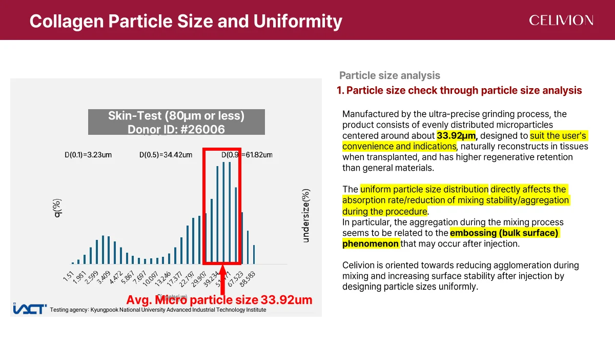
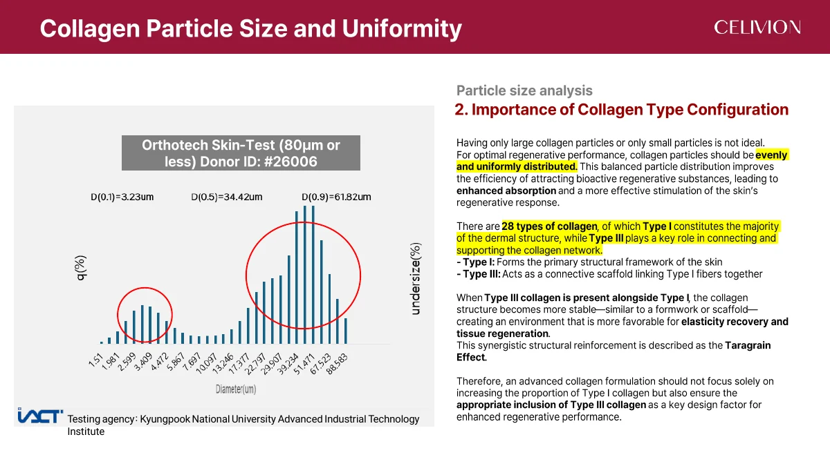
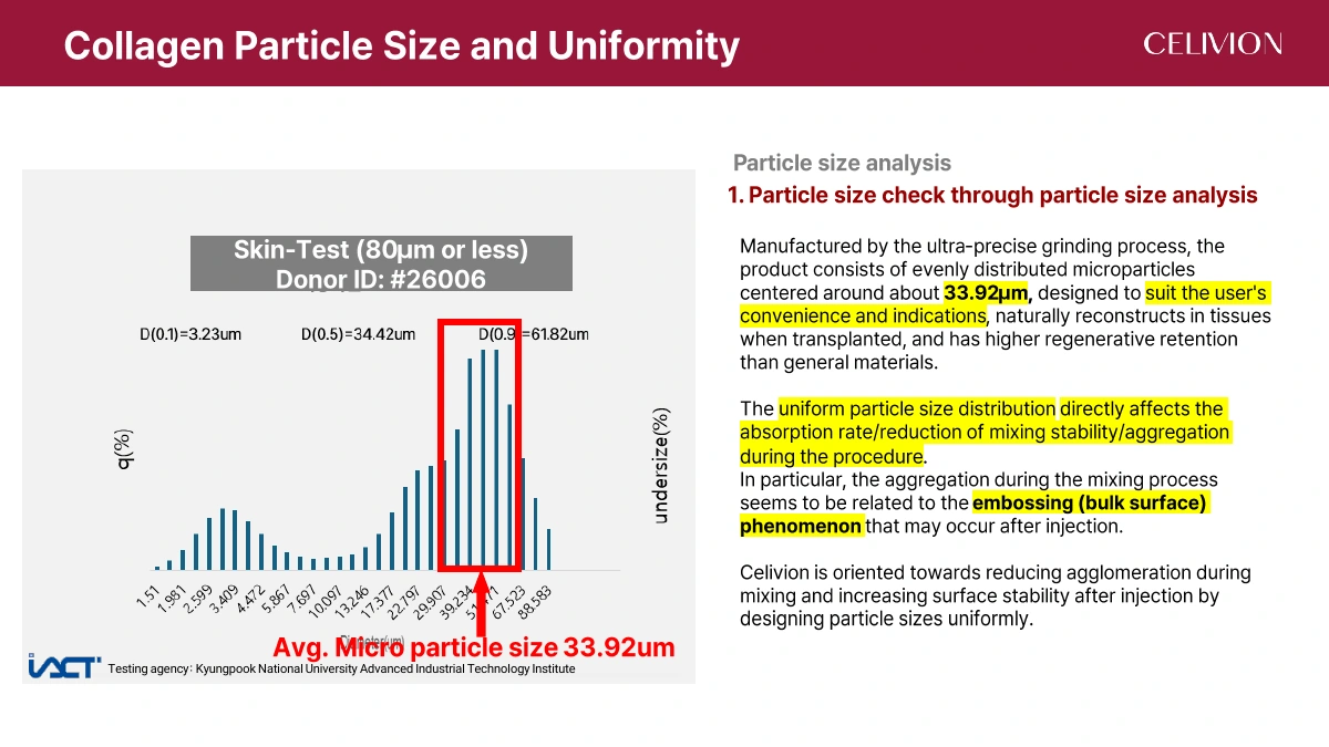
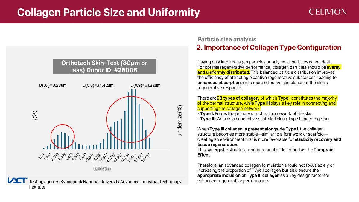
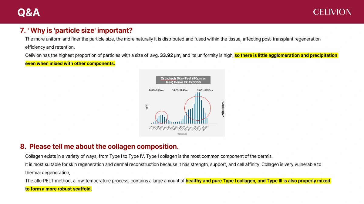
The Celivion Lin J dossier reports an average particle size of 33.92 μm. Its chart is labelled “Skin-Test (80 μm or less)” and donor ID #26006. Testing institution: Kyungpook National University Advanced Industrial Technology Institute.
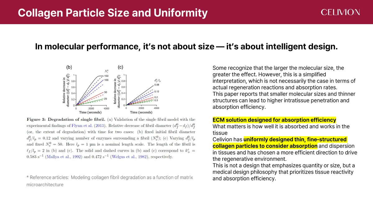
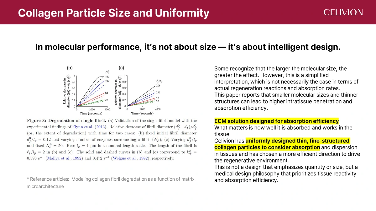
The source discusses particle uniformity in relation to dispersion and agglomeration. It cites “Modeling collagen fibril degradation as a function of matrix microarchitecture” to introduce the role of architecture; that paper is not presented as a Celivion clinical trial.
The 33.92 μm value belongs to this dossier. Other source documents on the site have their own sample values and measurement contexts; they are not substituted for this result.
Average particle size
Celivion Lin J · #26006
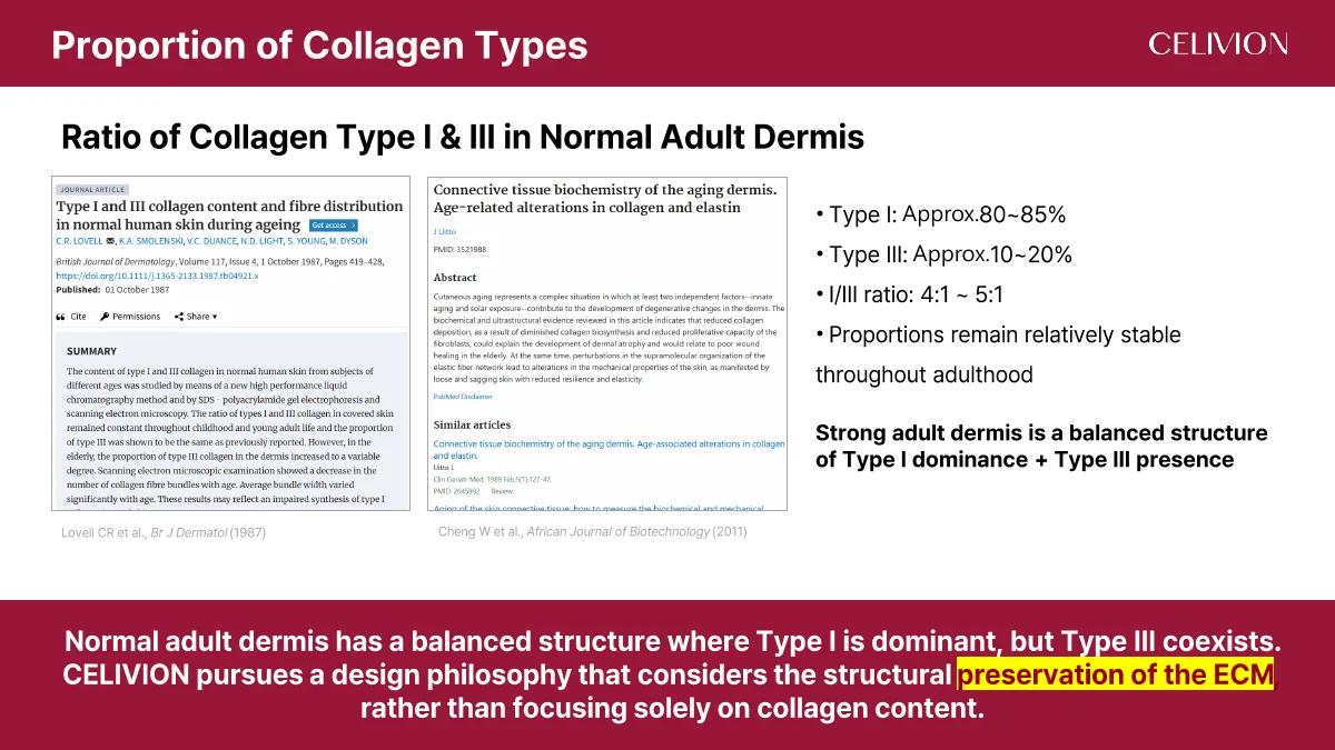
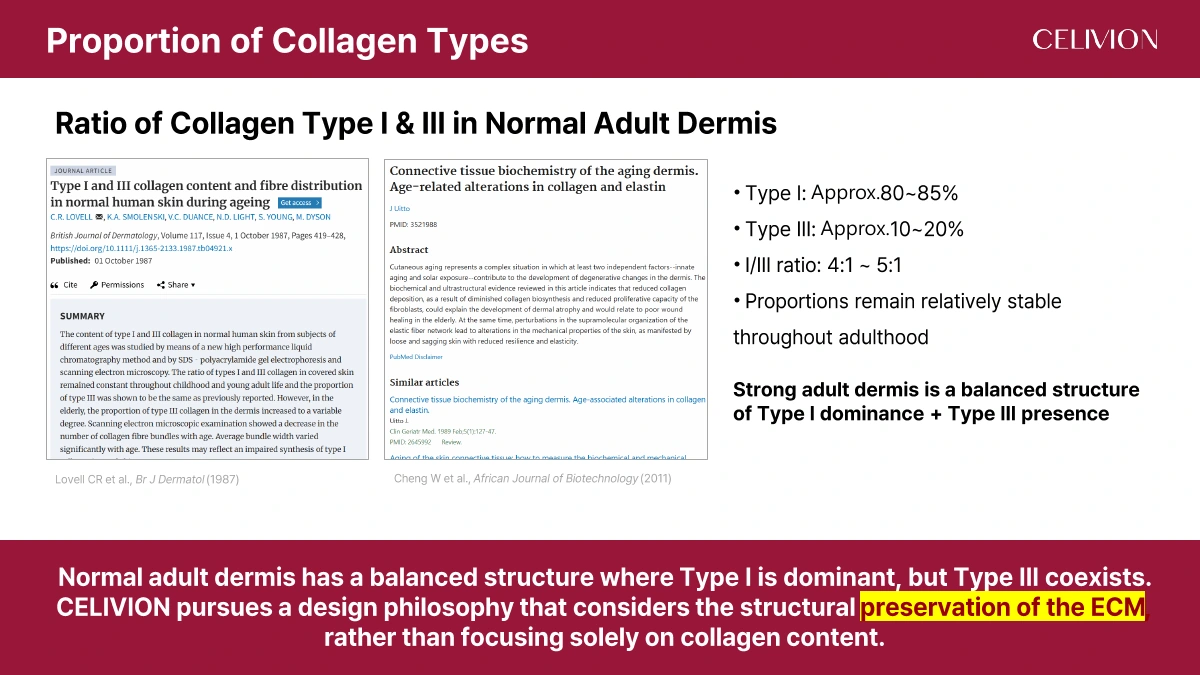
The source describes adult dermal collagen as predominantly Type I (approximately 80–85%), with Type III (approximately 10–20%) and an I/III ratio of about 4:1–5:1. Type I provides structural support; Type III contributes to the supporting network. These are background dermal values, not measured percentages for Celivion Lin J.
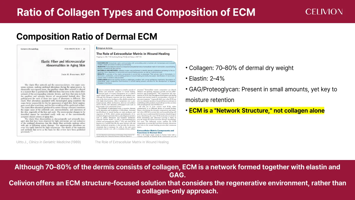
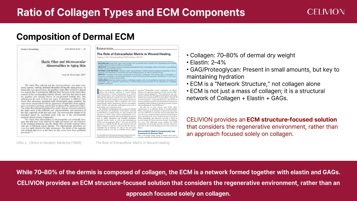
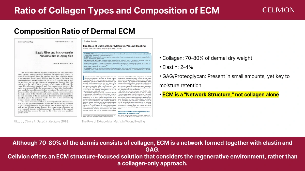
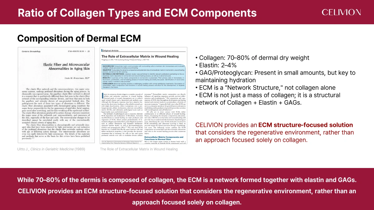
The dossier lists collagen at 70–80% of dermal dry weight, elastin at 2–4%, and smaller amounts of glycosaminoglycans (GAGs) and proteoglycans involved in hydration. These general dermal composition ranges are separate from the product analysis above.
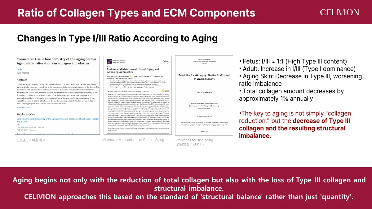
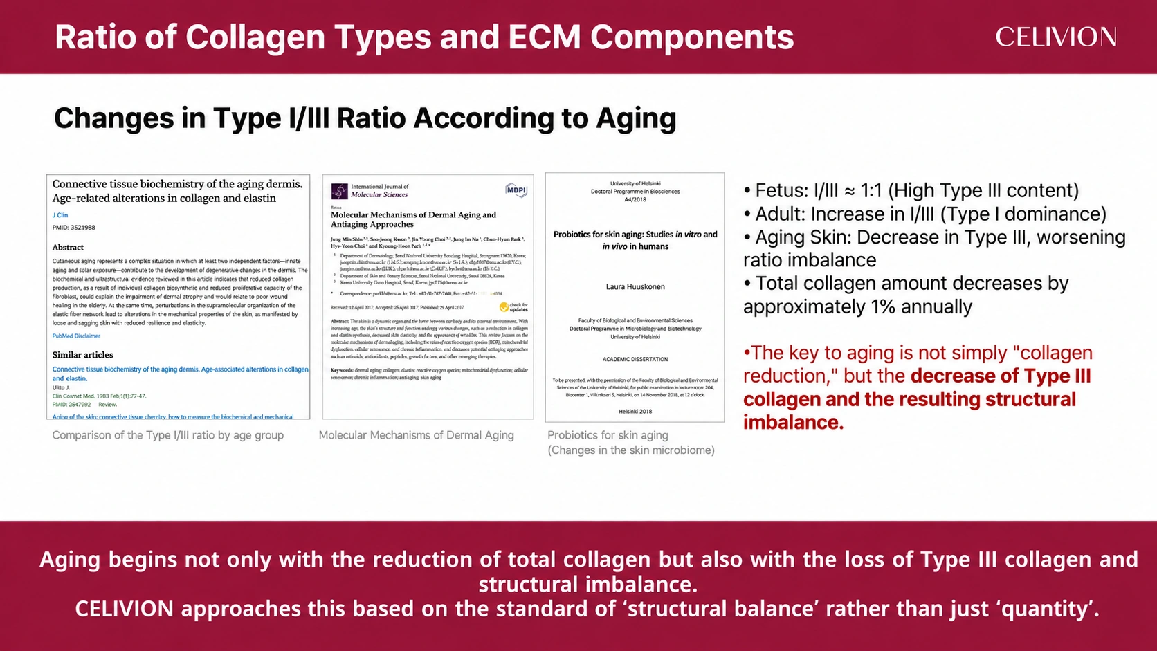
For ageing, the source discusses changes in Type I/III balance, a fetal ratio near 1:1 and an approximate annual collagen reduction of 1%. Its design narrative focuses on preserving ECM structure rather than collagen quantity alone.
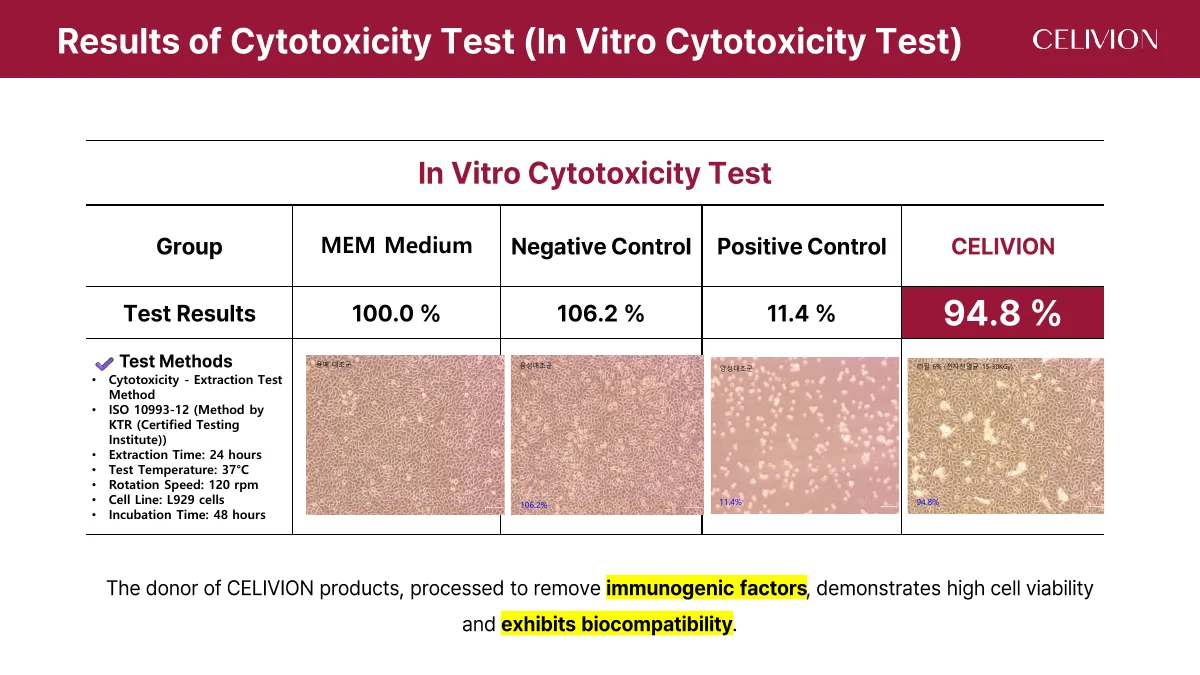
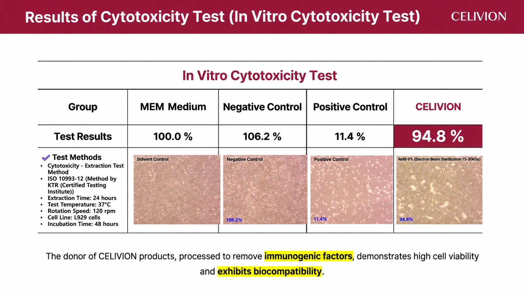
The in vitro cytotoxicity table reports 94.8% for Celivion, compared with 100.0% for MEM medium, 106.2% for the negative control and 11.4% for the positive control. This is a cell viability result in the stated test, not a clinical success rate.
The source lists an extraction test, ISO 10993-12, KTR, extraction for 24 hours at 37°C and 120 rpm, L929 cells and incubation for 48 hours.
| Group | Cell viability |
|---|---|
| MEM medium | 100.0% |
| Negative control | 106.2% |
| Positive control | 11.4% |
| Celivion | 94.8% |
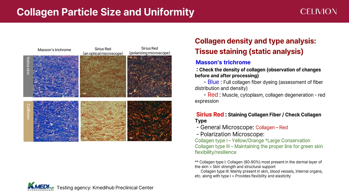
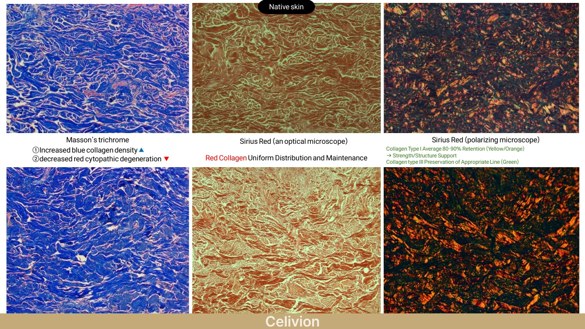
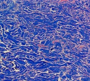
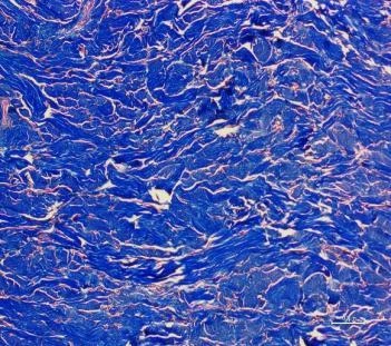
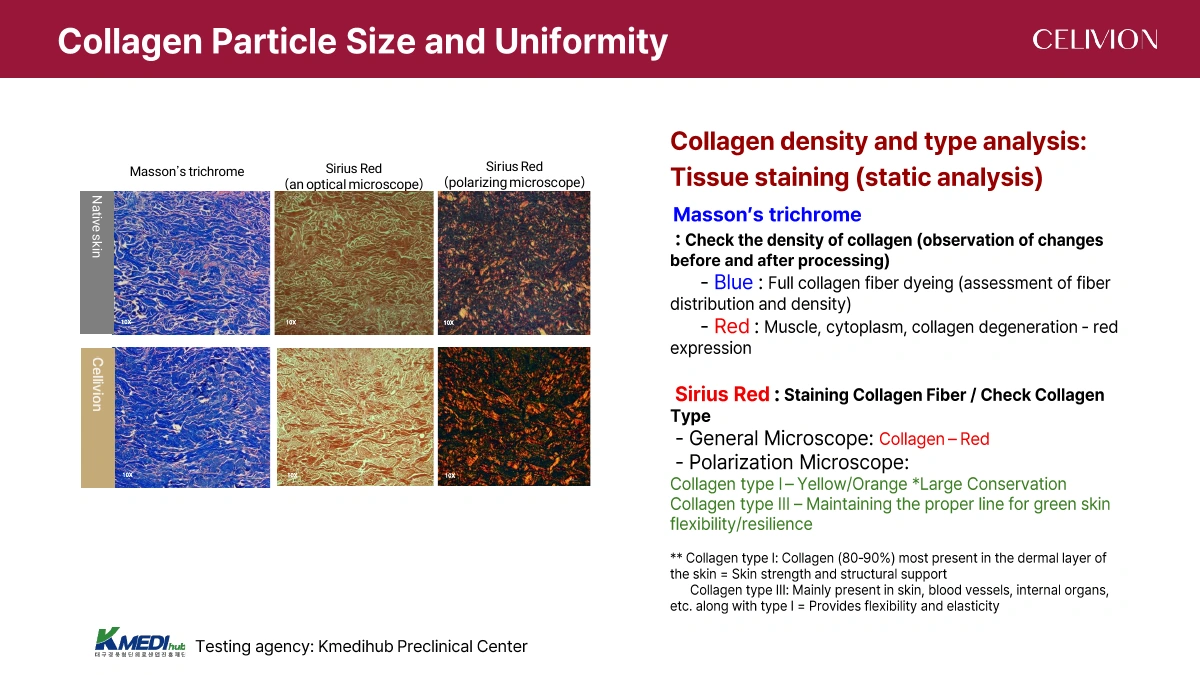
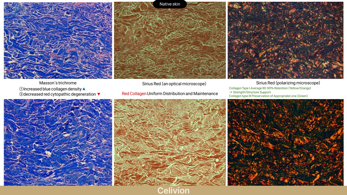
Masson’s trichrome is used to compare collagen fibre distribution and density before and after processing. The source identifies blue with collagen fibres and describes red staining of other tissue components.
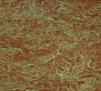
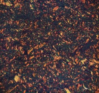
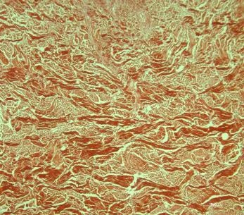
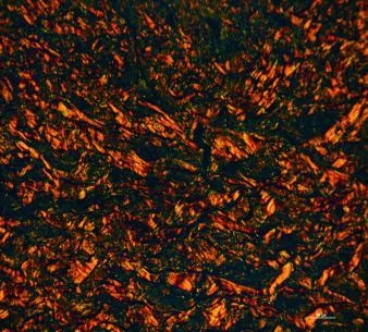
Sirius Red images are shown under optical and polarizing microscopy. The dossier associates yellow/orange with Type I and green with Type III in its interpretation of the images. The panels compare native skin and Celivion at 10×.
The original microscopy images are retained without recolouring. Testing institution named in the source: Kmedihub Preclinical Center.
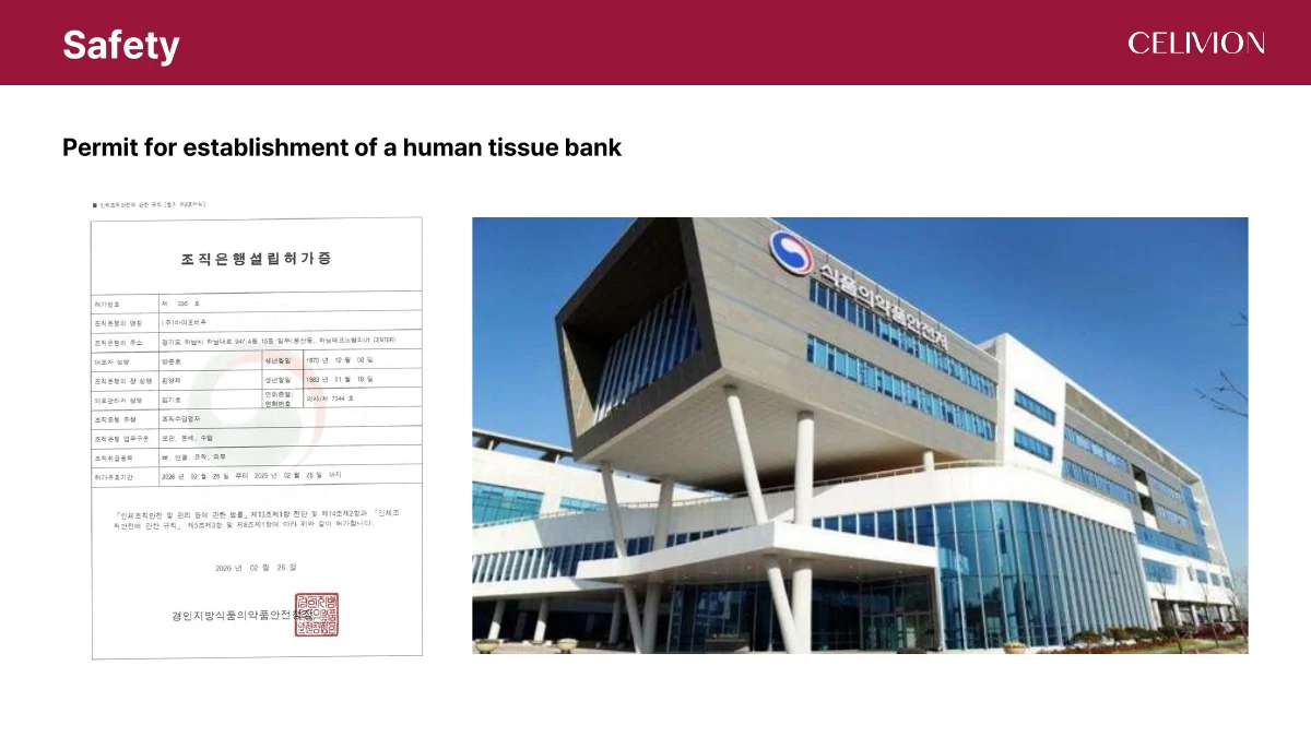
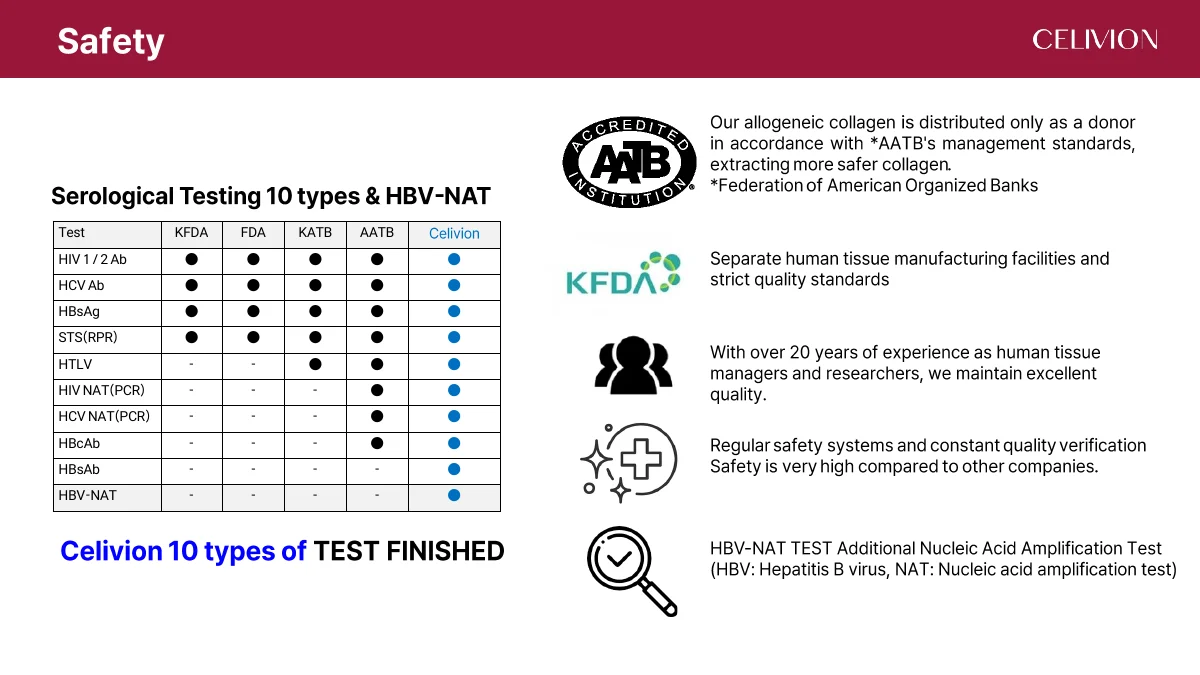
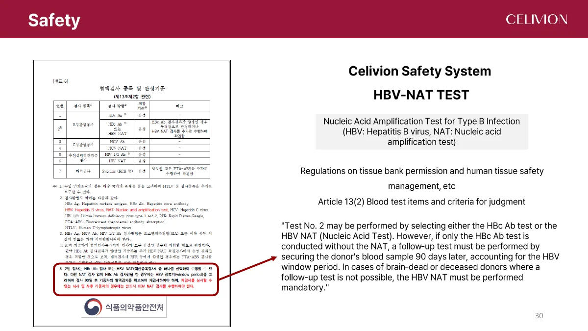
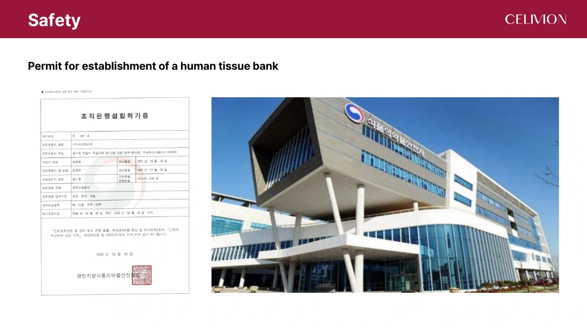
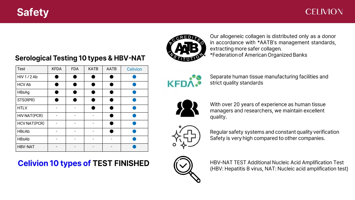
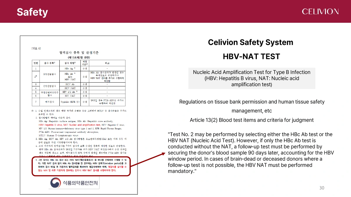
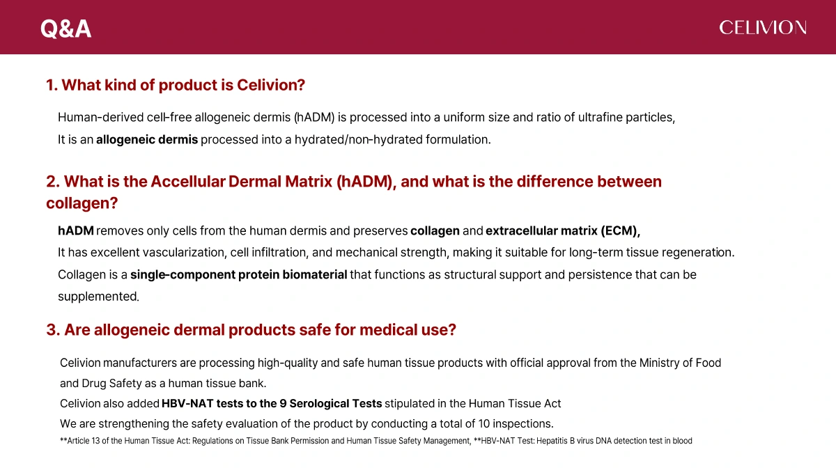
The dossier presents a Korean human tissue-bank establishment permit and describes donor screening, dedicated tissue processing facilities and quality verification. Its screening list contains ten entries, including HBV-NAT.
AATB accreditation concerns tissue establishments and their specified activities. References to AATB in the source are not presented here as approval of a Celivion Lin J aesthetic indication.
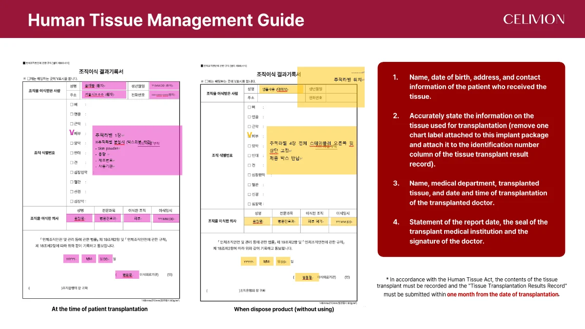
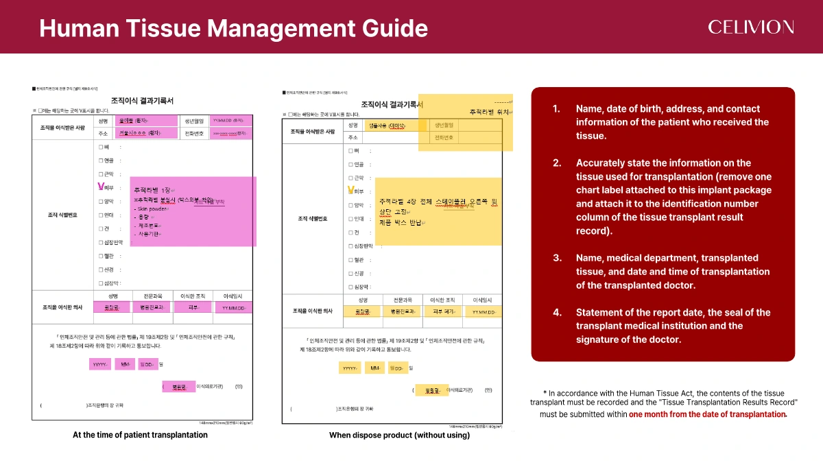
The source’s Korean tissue-management guidance covers recipient identification, tissue identification labels, clinician and procedure records, institutional confirmation and reporting within one month. This describes the source’s Korean framework, not a universal rule for every country.
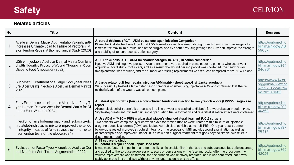
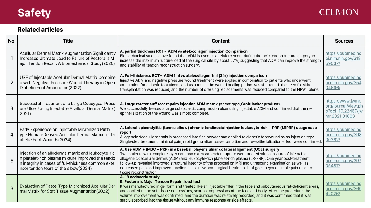
The dossier cites research on ADM in tendon repair, wound care and soft-tissue augmentation. The publications below concern their own materials, populations and study designs; they are not all studies of Celivion Lin J. Titles and identifiers are retained for direct retrieval.
The complete 37-page English dossier is available below. Web sections consolidate related pages; each section links to its source pages.
PDF 33–37
Based on the dossier’s questions, pp. 33–37.
A product based on human acellular dermal matrix (hADM), processed into fine particles. The technical dossier describes its material structure, processing and laboratory results.
hADM retains a dermal ECM network after cell removal. Collagen is a protein component within that network; the terms do not describe the same material composition.
Donor screening, ten listed infection-screening entries including HBV-NAT, residual DNA, tissue staining, surfactant residue and in vitro cytotoxicity. Each test addresses a specific endpoint.
The source describes staged low-temperature circulation, preservation of ECM components and particle sizing. It states that acetic acid and pepsin are not used.
Temperature affects collagen structure. The dossier presents low-temperature processing as a way to limit thermal deformation while removing cellular material.
It is the residual DNA value reported for Celivion in this dossier. It informs assessment of decellularization, alongside other tests; it is not a zero-risk guarantee.
The reported average is 33.92 μm. The source chart identifies donor #26006 and a Skin-Test sample labelled 80 μm or less.
Types I and III are discussed in relation to dermal support and network balance. General adult-skin ratios are background information, not a product-specific assay.
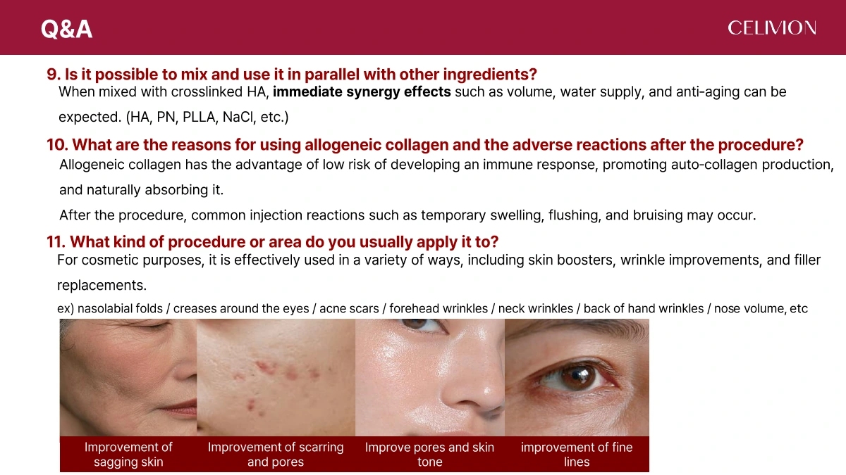
Page 37 mentions HA, PN, PLLA and NaCl. It does not provide a validated compatibility protocol for each combination. This page documents that topic and does not prescribe mixing or administration.
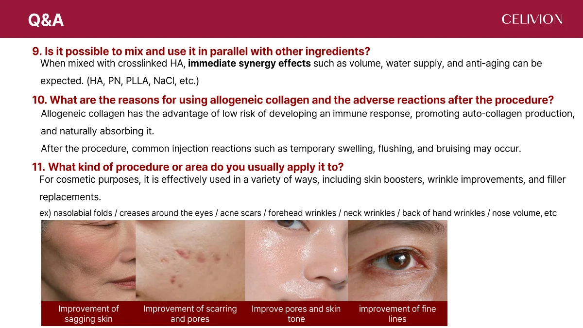
Temporary swelling, flushing and bruising are listed. The source’s list is not a complete adverse-event profile.
The dossier discusses skin texture, pores, fine lines, scars and tissue support. These are source topics, not a list of approved indications in every jurisdiction.
CELIVION · SOURCE LIBRARY
Figures follow the selected page language. Product packaging, original test photographs, certificates, forms and published-paper facsimiles retain their original appearance. The original English PDF remains available for verification.
Read the source PDF · 37 pages ↗ Page 01 ↗
 Page 02 ↗
 Page 03 ↗
 Page 04 ↗
 Page 05 ↗
 Page 06 ↗
 Page 07 ↗
 Page 08 ↗
 Page 09 ↗
 Page 10 ↗
 Page 11 ↗
 Page 12 ↗
 Page 13 ↗
 Page 14 ↗
 Page 15 ↗
 Page 16 ↗
 Page 17 ↗
 Page 18 ↗
 Page 19 ↗
 Page 20 ↗
 Page 21 ↗
 Page 22 ↗
 Page 23 ↗
 Page 24 ↗
 Page 25 ↗
 Page 26 ↗
 Page 27 ↗
 Page 28 ↗
 Page 29 ↗
 Page 30 ↗
 Page 31 ↗
 Page 32 ↗
 Page 33 ↗
 Page 34 ↗
 Page 35 ↗
 Page 36 ↗
 Page 37 ↗